The Kathmandu Earthquake will Alter Brain Structure of Survivors
- Published26 Apr 2015
- Author Douglas Fields
- Source BrainFacts/SfN
The disastrous earthquake in Kathmandu has killed hundreds of people and brought grievous tragedy to thousands. Even among the survivors, the earthquake will leave its mark in the form of altered brain structure, according to neuroimaging research performed on survivors of the Wenchuan earthquake of 2008. Studies by Lui and colleagues on survivors of the 2008 Wenchuan earthquake in China report changes in brain structure that can be seen by MRI. The 7.9 magnitude Wenchuan earthquake (also called Sichuan earthquake) rocked the mountainous central region of Sichuan province in southwestern China on May 12, 2008. 90,000 people died. 375,000 people were injured. Millions of people were rendered homeless.
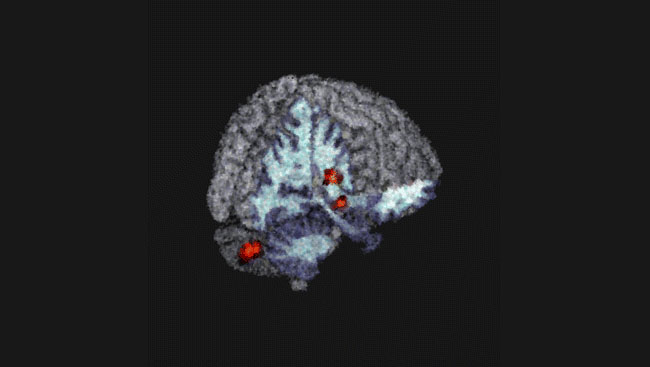
A 2013 study was performed on 44 survivors, male and female, 25 days after the earthquake and compared to 38 matched controls who had their brains scanned for other reasons prior to the earthquake. The results showed a decrease in gray matter in the insula, hippocampus, and caudate, and an increase in the orpitofrontal cortex (OFC) and parietal cortex.
The OFC is important in modulating emotional responses in the hippocampus, amygdala, ventral striatum and insula. The increase in gray matter is consistent with elevated demands for top-down (that is executive functioning of the cerebral cortex) regulation of threat, fear, and stress circuitry in the limbic system. The increase in parietal cortex has been reported previously in other types of trauma survivors, and this may reflect enhanced neural activity related to the hyper-vigilant state.
The authors suggest that the lower grey matter volume in the insula, striatum, and hippocampus may result from decreased neurogenesis and increased synapse elimination that are seen in studies of experimental animals subjected to chronically elevated stress hormone levels. Acute stress elevates corticotropin-releasing hormone to activate the hypothalamic-pituitary-adrenal axis that is engaged in the “fight-or-flight” response, but prolonged elevation of this stress response is damaging to the brain and body in many respects.
Scientific data on the effects of stress on the human brain are difficult to obtain for ethical reasons, and extrapolating complex cognitive processes of human stresses from animal research is problematic. Studies of people who have survived natural disasters or traumatic events can provide important insights into the effect of stress on human brain structure.
Similar brain changes have been observed in people who have experienced other major life stresses. Studies have reported altered brain structure in patients with PTSD involving regions that function in threat detection and fear, notably the amygdala, hippocampus, and prefrontal cortex (anterior cingulate and medial frontal gyrus). The altered gray matter volume in the prefrontal to limbic and striatal systems found in earthquake survivors are recognized to be involved in emotional and conscious decision making. The striatum and parietal regions are activated in making decisions under time pressure. These regions also undergo changes in people with anxiety disorders, and these brain regions are engaged when processing fear and pain.
The authors conclude that survivors of severe emotional trauma may experience substantial change in brain function and also in the structural anatomy of the prefrontal-limbic, parietal and striatal brain system. These changes are not necessarily pathological. Rather they reflect in part the brain’s remarkable capacity to modify its structure and function rapidly in response to environmental experience. The changes found in earthquake survivor’s brains likely increase the ability of survivors to respond rapidly and appropriately to the danger and trauma. However, if these brain modifications do not return to normal after the threat has passed, this can result in dysfunction. This is well demonstrated by brave military men and women who suffer post-traumatic stress disorder after returning to a safe environment. Similar changes in the brains of people under stress that helped them survive in combat, can become debilitating after returning to civilian life.
About the Author

R. Douglas Fields is Chief of the Nervous System Development and Plasticity Section at the National Institutes of Health, NICHD, in Bethesda, Maryland, and author of the new book about sudden anger and aggression “Why We Snap,” published by Dutton, and a popular book about glia “The Other Brain” published by Simon and Schuster.
CONTENT PROVIDED BY
BrainFacts/SfN
References
Cohen, R.A. et al., (2006) Early life stress and morphometry of the adult anterior cingulate cortex and caudate nuclei. Biol. Psychiatry 59: 975-82.
Davidson, R.J. (2000) Dysfunction in the neural circuitry of emotion regulation -- a possible prelude to violence. Science 289: 591.
Lui, S. (2009) High-field MRI reveals an acute impact on brain function in survivors of the magnitude 8.0 earthquake in China. Proc. Natl. Acad. Sci. USA 106; 15412-7.
Lui, S., et al., Bran structural plasticity in survivors of a major earthquake. J Psychiatry Neurosci. 2013 Nov;38(6):381-7
McEwen, B.S. (2007) Physiology and neurobiology of stress and adaptation: control of the brain. Physiol. Rev. 87:873-904.
Rafferty, J. P. (2008) Sichuan earthquake of 2008. Encyclopedia Britannica.
Also In Anatomy


Trending
Popular articles on BrainFacts.org