Neurons: A Curious Collection of Shapes and Sizes
- Published22 Aug 2012
- Reviewed22 Aug 2012
- Author Susan Perry
- Source BrainFacts/SfN
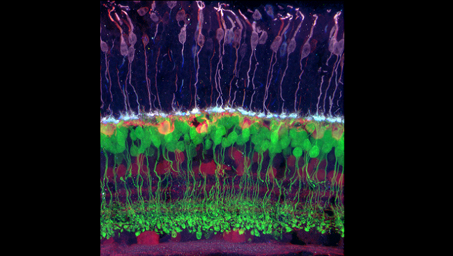
Neurons — the nerve cells that make up the brain and nervous system — look different from all other cells in the body. And from one another.
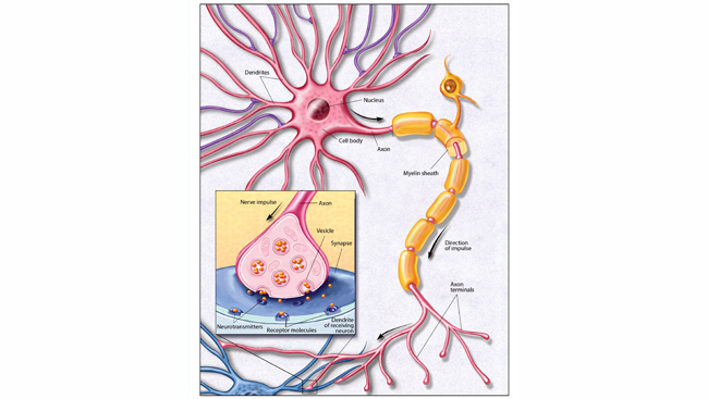
Like blood, liver, muscle, and other body cells, neurons have an outer membrane, a nucleus, and smaller structures called organelles that perform important functions. But neurons also have something other cells don’t: highly complex extensions called dendrites and axons that transport electrical and chemical messages in and out of the cell, enabling neurons to communicate with one another with incredible speed and precision.
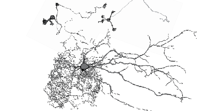
The intricate branches, or arbors, of these extensions are what give neurons their beautifully strange and varied shapes. Dendrite arbors, for example, make some neurons look like sea coral, others like spider webs, and still others like round balls of tumbleweed. Axonal arbors are equally diverse. They can have a simple T shape and be quite short (less than one inch). Or they can be multi-branched and stretch, as do the axons of the sciatic nerve that run along the back of the thigh, for as long as three feet. By studying the structural diversity of neurons, scientists are gaining greater insight into how the brain and the rest of the nervous system works.
An extraordinary diversity
Scientists have been identifying and classifying neurons for more than 100 years. The 19th-century Spanish physician and pathologist Santiago Ramón y Cajal discovered that the brain and the rest of the nervous system consisted not of one jumbled mass of tissue, but of discrete cells. Cajal’s exquisitely detailed drawings of neurons provided scientists with the first evidence of their structural diversity.
That diversity is extraordinary. Scientists have identified hundreds of types and subtypes of neurons using advanced cellular imaging techniques, and more are discovered each year.
“In a sense, neurons are like snowflakes,” says Kristen Harris, a neurobiologist at the University of Texas who studies how changes in neuron structure affect learning and memory. “They have recognizable shapes, but no two look exactly alike.”
But is there, as Cajal proposed a century ago, a unifying theory behind the diversity of the neuronal shapes? Do neurons form their shapes in a predictable way and for specific purposes?
Surprising findings
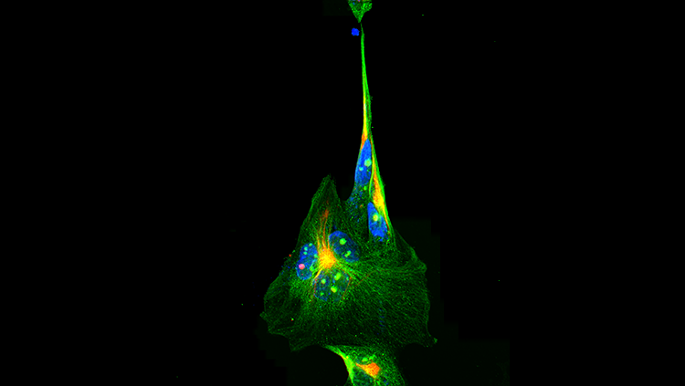
Recent discoveries enable scientists to come closer to answering these and other questions about the shapes of neurons. The investigation into how axons and dendrites form (and thus determine a neuron’s shape) has revealed a surprising finding: Although axons form by growing to their target location like plants growing toward sunlight, some dendrites develop by a process called retrograde extension — whereby the cell anchors its dendrite tips at the target and then pulls back, away from the target.
Neurons do not appear in isolation, but are organized into bigger shapes called neuropils, dense regions of axons, dendrites, and synapses (the tiny information-transferring gaps between neurons). Recently, Harris, in collaboration with neuroscientist Terrence Sejnowski, at the Salk Institute for Biological Studies, and others, created a 3-D computer model of a small volume of neuropil from the brain of a rat.
“The result is a puzzle that we can take apart and put back together in the computer,” Sejnowski says. The model has already revealed that the spacing between neurons is not uniform and that the ends of dendrites and their spiny protrusions vary enormously in size. What this variety means in terms of brain function is not yet known.
A way to save space
Scientists have also learned that neurons are not placed helter-skelter in the brain, even though their placement often looks random. Careful measurements have shown that neurons are organized in space to minimize the lengths of the axons needed to connect them — possibly to pack more neurons into the brain and thus increase its processing capability.
“Form and function has been fundamental to the discovery of how things work in the nervous system,” Harris says. “But no neuron has just one function. We’ve learned a great deal about the diversity and complexity of neuronal shapes, but we have much, much more left to discover.”
CONTENT PROVIDED BY
BrainFacts/SfN
References
Patel MR, Shen K. Neurite extension: starting at the finish line. Cell. 2009;137(2):207-209.
Stevens CF. Brain organization: wiring economy works for the large and small. Current Biology. 2012;22(1):R24-5.


