Brains are made of plastic. Seems like a ridiculous claim. The brain is not made of plastic. Or, is it? In neuroscience we often refer to the brain as being plastic, but, what is plastic? The definition that most of us are familiar with is the material known as plastic. Plastic can come in many shapes and sizes like you see below.

These are some common household examples of plastic and what’s important to note here is that plastics are highly malleable. Constructed from primarily from organic polymers, these chemicals can be constructed to serve a variety of functions and in some cases can conduct electricity. Plastics can be molded to make rigid fibers, or something elastic, like rubber.
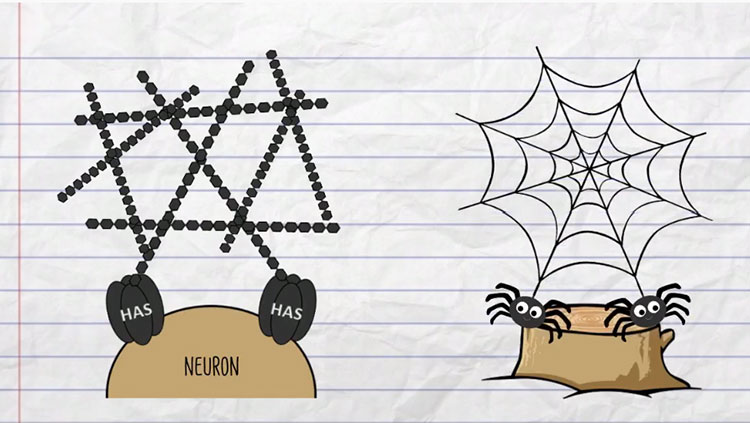
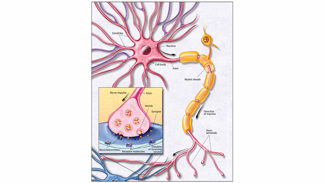
Why would we call the brain plastic? Well, if we can use our imagination for a moment and say that neurons and glia are the polymers of the brain, then indeed, there are some very straightforward comparisons. Collections of neurons linked together can indeed conduct electricity. The lipoprotein myelin insulates the axon of neurons which act as wires and allow for the rapid conduction of electricity across great distances within organisms. In giraffes, the cell body and axon of neurons composing the recurrent laryngeal nerve span 15 feet!
While these corollaries to plastic are interesting, they are not what neuroscientists mean when we use that term. And we use that term a lot.
What we mean by, “The brain is plastic,” is that the brain has a unique ability – the ability to change. The brain is not a static organ. Given our fantastic capacity for learning and remembering, it might not be much of a stretch for us to accept that brain functions can change over time. And, as a side note, plastic is a material, whereas plasticity is the ability of a material to be altered. So in the case of neuroscience, the nervous system is the material that displays plasticity, or the ability to undergo change.
As someone that works in a physiology lab, whenever I use or hear that term, I am biased. I always think about plasticity in terms of brain of function, or alterations in neuronal or neural network function as measured by electrophysiological techniques. I would be amiss though if I didn’t mention that there are many forms of plasticity in the brain. Spines on the dendrites of neurons can disappear or grow, synapses can increase in number, protein expression in neurons or glia can be altered, and behaviors can change. There are quite a few examples of these plastic changes throughout the entire central and peripheral nervous system.
As I am currently working on a project involving measuring alterations in long-term potentiation (LTP; a physiological type of plasticity), I find myself fixated on this concept. Most people in neuroscience know it as the proposed mechanism of learning and memory. In the most basic terms, long-term potentiation is a lasting increase in the strength of communication between specific neurons at specific synapses. Another way of saying this is that before LTP, you have a response of magnitude x from a given neuron or network when using stimulation y. Then, after one of numerous methods of inducing LTP, you have an increase in the magnitude of the response x using the same stimulation value y. For an example of this, see the below figure from the Wikipedia article on long-term potentiation.
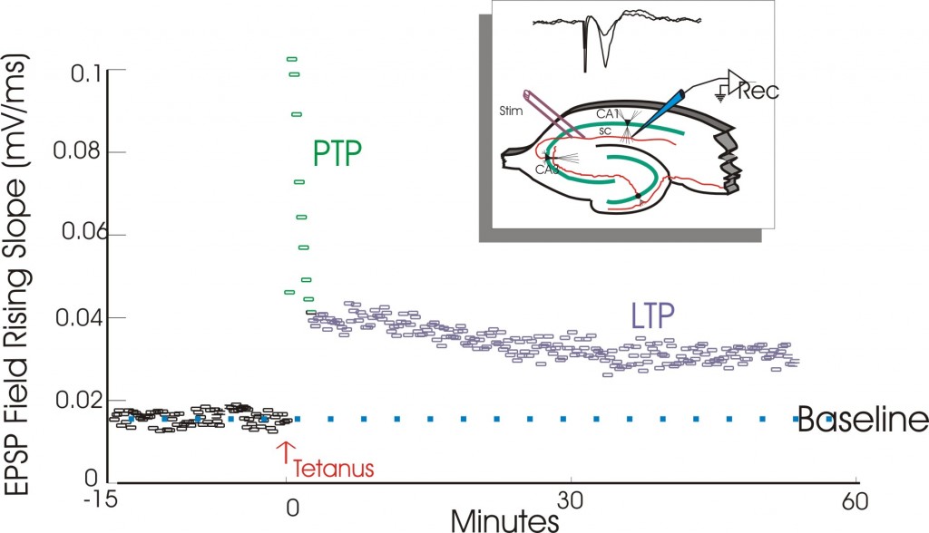
This is a classic example of a physiological recording conducted in the hippocampus, a key brain region involved in learning and memory. Although almost all synapses in the brain probably display plasticity, the hippocampus is very commonly studied for LTP as it displays extremely robust LTP that is not only easy to elicit experimentally but is fairly large in magnitude. In these experiments, you stimulate the input (axons) that synapse onto a network of cells in an area of the hippocampus referred to as Cornu Ammonis 1 (or CA1). Then, with your recording electrode you measure a baseline response following repeated stimulation of the same stimulation magnitude (each data point on the figure above represents the slope of the response seen on the inset figure above the electrode placement diagram) and then deliver a rapid burst of stimulation (referred to as a tetanus). Following a tetanus, CA1’s network response is enhanced when the stimulation is the same as it was before the tetanus was delivered. Now, what does all of this mean? If this post-tetanus increase in CA1's response persists over time (typically at least 1 hour), then LTP has been induced.
Another characteristic of this physiological concept is that it is specific to the synapse studied. An abstract example of this might be if you are learning calculus. Imagine you have a synapse in your brain that specifically responds to learning how to do integration. Once you learn it, this memory persists (maybe at least during lecture that day) and is theoretically specific to that synapse. If we were able to record from this synapse, we might observe LTP during the lecture when you were learning integration. Now, does this mean we have a blank synapse waiting around that has been pre-assigned to learning integration? Probably not. But, like free memory space in a computer, we have new synapses forming every day and these are probably sequestered for the storage of new memories and/or for learning. What’s amazing about the plastic nature of the brain is that these changes are–in most cases–reversible.
Another physiological concept that in some ways mirrors LTP is known as long-term depression (or LTD). Similar to the LTP figure above, you would have a baseline response as your 100% or average, stable response and then in the case of LTD, after a different stimulation paradigm you can depress this response to be less than 100% persistently.
Deficits in the ability of certain synapses to undergo LTP/LTD may underlie many different disorders. For example, in a recent report by Isono and colleagues in Neuroscience Research, they found that a fragment of amyloid-β (a protein that is found deposited in the brains of Alzheimer’s patients) disrupted LTP in the hippocampus of mice. Since the hippocampus is critical for the formation, and the recall of long-term memories, this impairment in LTP may underlie the hallmark memory deficits observed in Alzheimer’s disease.
The brain is obviously not made of plastic. But, I hope after reading this you’ve learned a basic property of the brain–of your brain: plasticity. Lastly, I want to mention how this was all discovered and how the field of studying plasticity was sparked because I find it to be fascinating. I used to think that the age-old idiom, “It’s all about who you know,” only had some truth to it, but when it came to the discovery of LTP, this phrase was certainly true. Without meeting Per Andersen in the streets of Oslo, Norway, Terje Lømo may have never conducted the first experiments eliciting LTP in the rabbit hippocampus.
And, in his short review recalling the discovery of this phenomenon, he is unclear how or when this physiological property of the brain would’ve been discovered without their serendipitous meeting.
About the Author

Steven Miller is a Ph.D. candidate at the Uniformed Services University of the Health Sciences in Bethesda, Maryland. Steven’s research focuses on identifying treatments against seizures induced by the chemical weapons known as nerve agents.
CONTENT PROVIDED BY
BrainFacts/SfN