Cushioning the Brain
- Published9 Oct 2015
- Reviewed7 Oct 2015
- Author Alexis Wnuk
- Source BrainFacts/SfN
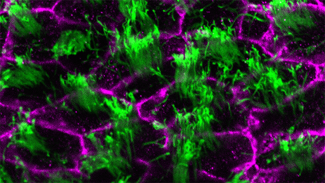
In addition to the skull and connective tissue, the brain is also protected by a cushioning fluid called cerebrospinal fluid (CSF). This fluid circulates through the cavities of the brain called ventricles, and into the space outside the brain and spinal cord before being absorbed into the bloodstream. This image shows the cells (purple) lining the ventricles in the brain of an adult mouse. These cells have tiny hair-like projections called cilia (green) that beat in the same direction to enable the flow of CSF. However, defects in the positioning of cilia can lead to problems with the flow of CSF and can cause hydrocephalus, a dangerous neurological condition where fluid accumulates in and around the brain. This can cause the ventricles to expand, putting pressure on the brain. By studying what causes cilia to develop abnormally, scientists hope to improve the diagnosis and treatment of hydrocephalus.
CONTENT PROVIDED BY
BrainFacts/SfN


