Understanding Involuntary Movements
- Reviewed5 Oct 2022
- Author Karen Hopkin
- Source BrainFacts/SfN
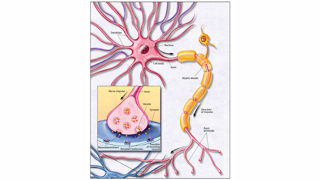
Voluntary movements like running and dancing may get all the attention, but involuntary movements, which take place without our conscious control, play a crucial role in everyday life. Among the simplest and most fundamental types of involuntary movements are the reflexes. Reflexes are relatively stereotyped, automatic muscle responses to particular stimuli — think of the rapid withdrawal of your hand after touching something hot. These reflexes involve the activation of sensory receptors in the skin, the joints, or even in the muscles themselves. The responses are rapid and occur without involvement of the brain or conscious attention. Instead, they depend on circuits of neurons located in or near the spinal cord itself.
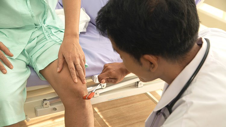
One of the best-known reflexes is the “knee jerk” response, a stretch (myotatic) reflex that occurs when a physician strikes the tendon just below the knee with a small rubber hammer. This tap produces a slight stretch of the knee extensor muscle, which is “sensed” by receptors within the muscle called muscle spindles. The spindles sense the extent and speed of the stretch, and stimulate sensory neurons, which send a barrage of impulses into the spinal cord. There, the signals activate the alpha motor neurons that cause the stretched extensor muscle to contract, triggering the reflex.
Of course, for the leg to kick forward, the antagonist flexor muscle has to relax at the same time. In fact, the same sensory stimulus that directly activates the motor neurons controlling the extensor also indirectly inhibits the motor neurons controlling the antagonist flexor. This reciprocal inhibition is accomplished by connecting neurons that lie completely within the spinal cord. When these so-called inhibitory interneurons are activated by the original sensory stimulus, they send impulses that inhibit the motor neurons supplying the flexor. Thus, even the simplest of reflexes involves the synchronous activation (and inactivation) of multiple sets of motor neurons controlling both agonist and antagonist muscles.
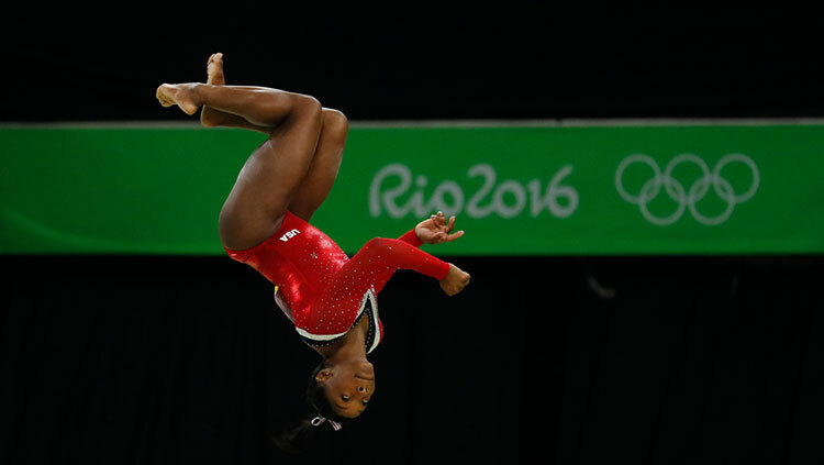
Many reflexes protect you from injury. When you’re seated in a doctor’s office, the “knee jerk” reflex simply makes your lower leg swing briefly forward. However, if you were to jump off a chair (or perform an even more dramatic gymnastic dismount) this same reflex would promote the contraction of the strong muscles that straighten your knees, helping you to “stick your landing” and remain upright.
Another protective reflex is the flexion withdrawal reflex that occurs when your bare foot encounters a sharp object. In this case, pain receptors in the skin send a message to the spinal cord, alpha motor neurons are activated, and the leg is immediately lifted (flexion). At the same time, because your body weight is supported on both legs, the extensors of the opposite leg must be activated. Without this additional reaction, called the flexion crossed extension reflex, you would lose your balance and fall over after stepping on a tack.
As all these movements occur, the muscles involved provide feedback to the brain with information about where the various body parts are in space and how fast they are moving. The muscle spindles mentioned earlier supply information about changes in muscle length or stretch. The brain, in turn, adjusts the sensitivity of the system via a separate set of motor neurons, called gamma motor neurons, which keep the muscle spindles taut. Other specialized receptors called Golgi tendon organs — located where the muscle fibers connect to the tendon — detect how much force or tension is applied to a muscle during ongoing movement, increasing the movement’s precision.
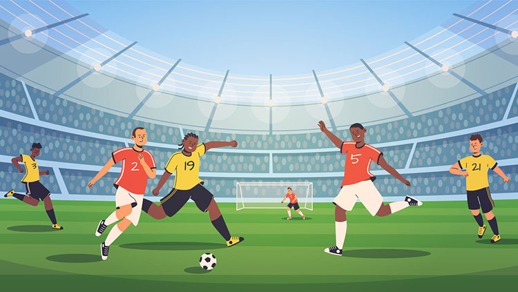
These feedback systems are not unique to reflexes but allow the brain to fine-tune how working muscles behave during a variety of movement tasks — from those that require a mastery of delicate positioning and coordination, such as sipping from a dangerously full teacup, to those that involve a targeted application of strength and speed, such as throwing a runner out at first base.
Adapted from the 8th edition of Brain Facts by Karen Hopkin.
About the Author

Karen Hopkin holds a PhD in biochemistry and is co-author of the textbook Essential Cell Biology.
CONTENT PROVIDED BY
BrainFacts/SfN
References
Ataxia. 2017. Diseases and Conditions. Mayo Foundation for Medical Education and Research. https://www.mayoclinic.org/diseases-conditions/ataxia/symptoms-causes/syc-20355652
Keihn, O., Dougherty, K., Pfaff, D.W. (ed.). 2013. Neuroscience in the 21st Century, Chapter 38, Springer Science+Business Media, LLC 2013. DOI: 10.1007/978-1-4614-1997-6_42
King, S. 2010. Reflex Reactions – Our Body’s Rapid Defense Mechanism. Positive Health Online. Issue 176. Compass Internet Ltd. http://www.positivehealth.com/article/chiropractic/reflex-reactions-our-body-s-rapid-defence-mechanism
Manto, M., Bower, J.M., Conforto, A.B. et al. 2012. Consensus Paper: Roles of the Cerebellum in Motor Control—The Diversity of Ideas on Cerebellar Involvement in Movement. Cerebellum 11, 457–487 (2012). https://doi.org/10.1007/s12311-011-0331-9
Marder, E., & Bucher, D. 2001. Central pattern generators and the control of rhythmic movements. Current biology: CB, 11(23), R986–R996. https://doi.org/10.1016/s0960-9822(01)00581-4
Shanmugarajah, P.D., Hoggard, N., Currie, S. et al. Alcohol-related cerebellar degeneration: not all down to toxicity? Cerebellum ataxias 3, 17 2016. https://doi.org/10.1186/s40673-016-0055-1
Wise S.P. and Shadmehr, R. 2002. Motor Control. Encyclopedia of the Human Brain. Vol 3.