Nuts and Bolts: The Neuron
- Published19 Jan 2012
- Reviewed24 Feb 2014
- Source Wellcome Trust
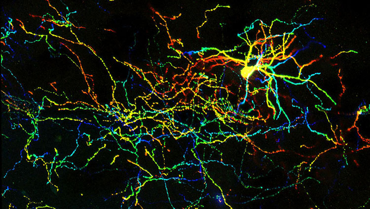
Neurons are highly specialised cells that conduct and process information in animals, enabling thought, perception and control of movement. Problems with neuronal function underpin a range of neurological and psychiatric disorders. Lydia Harriss presents a quick guide to these remarkable cells.
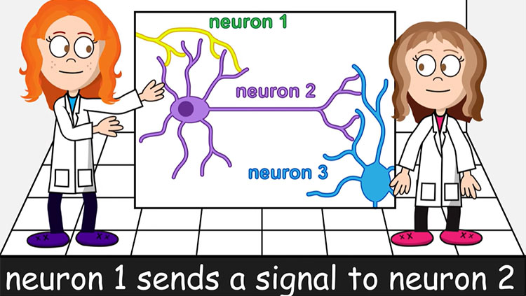
A single neuron may be connected to as many as 200 000 others, via junctions called synapses. They form an extensive network throughout the body, and can transmit signals at speeds of 100 metres per second. This enables animals to process and respond to events rapidly, for example by carrying sensory information from the ears to the brain, then instructions for movement from the brain to the leg muscles
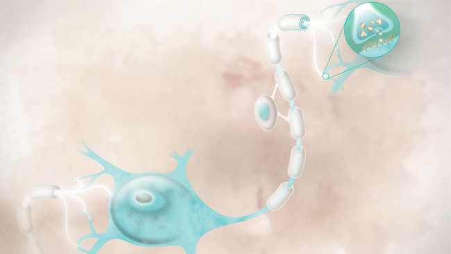
Within a neuron, signals are transmitted by a change of membrane voltage – a variation in the difference in electrical charge between the inside and outside of the cell. This electrical signal moves along the neuron as an electrical pulse (the ‘action potential’).
The nature of the connection between neurons was hotly debated until early-20th-century experiments by Otto Loewi and Sir Henry Dale (a founding trustee and chairman of the Wellcome Trust) showed that signals are typically transmitted across synapses by chemicals called neurotransmitters.
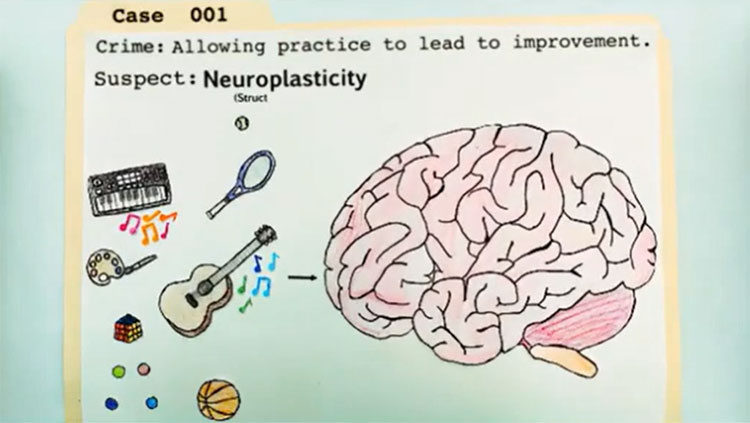
Researchers are investigating how changing levels of neuron activity alter the number of synapses and how well they transmit signals. This has given us insight into cognitive processes such as memory and learning, and has suggested treatments for diseases in which neural network activity becomes uncontrolled, such as epilepsy.
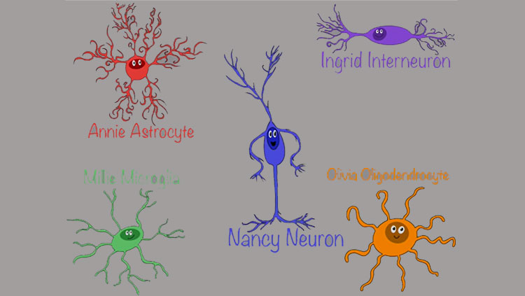
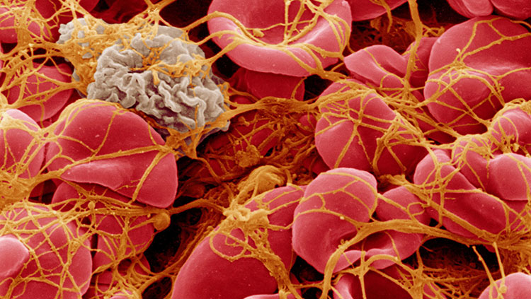
There is also great interest in glial cells, found in the spaces between neurons. Some glial cells (astrocytes) maintain the composition of this watery space, helping neurons to function properly. Others (oligodendrocytes) wrap neurons in an insulating myelin sheath, which can become damaged in neurodegenerative conditions such as stroke, spinal cord injury, multiple sclerosis and cerebral palsy. A better understanding of how neurons interact with glial cells may help in finding new treatments for these conditions
Nervous research
Current research in this field funded by the Wellcome Trust includes that of Professor David Attwell, University College London, who is investigating how proteins on the surface of certain glial cells may be responsible for the malfunction or death of neurons, as seen in conditions such as cerebral palsy, stroke and spinal cord injury.
Neurons can readily change, which allows them to adapt to variations in environment but also makes the networks that they form inherently unstable. Professor Juan Burrone, King’s College London, is studying how neurons avoid drifting towards extreme levels of activity. Understanding this better will provide targets for treating diseases caused by uncontrolled neuron activity, such as epilepsy.
Professor Peter Brophy, University of Edinburgh, has identified a gene that is mutated in people with a form of Charcot–Marie–Tooth disease, which affects the peripheral nerves outside of the brain and spinal cord. He is using mouse models to understand why the absence of the protein encoded by the gene makes peripheral nerves degenerate.
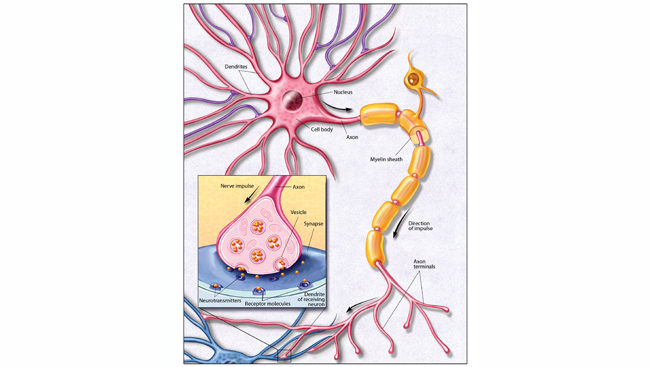
Parts of the neuron
Axon
The long projection that carries signals away from the cell body. The membrane voltage change from an incoming signal here triggers the opening of channels that allow ions (charged atoms) to flow into the cell from outside. This causes more channels farther along the axon to open, creating a voltage pulse that propagates along it.
Cell body (soma)
Contains many components typically found in other types of cell. This includes DNA, located in the nucleus, which holds instructions for producing the proteins that determine the shape and function of the cell.
Cell membrane
A film of fatty molecules that encloses the neuron.
Dendrites
Protrusions from the cell body that form branches connecting to other cells. These connections are input synapses, which receive signals from the axons of neighbouring neurons.
Myelin sheath
Many neurons are insulated by myelin: multiple layers of cell membrane that wrap around the axon. The sheath is interrupted at regular intervals (‘nodes of Ranvier’), where the channels that generate the electrical signal are located. Myelin reduces leakage of electrical charge from the axon, resulting in a signal that rapidly jumps from one node of Ranvier to the next, speeding up the conduction of information.
Oligodendrocyte
A type of glial cell that makes the myelin sheath.
Synapse
A connection between two neurons. When a nerve signal travelling along an axon reaches a synapse, it triggers the release of a chemical neurotransmitter that diffuses across the synaptic gap and binds to proteins on the surface of the receiving neuron. This binding causes an influx of ions, changing the membrane voltage and initiating an electrical signal in the second neuron.
CONTENT PROVIDED BY
Wellcome Trust