Mind Mapping
- Published10 Mar 2015
- Reviewed10 Mar 2015
- Author Alexis Wnuk
- Source BrainFacts/SfN
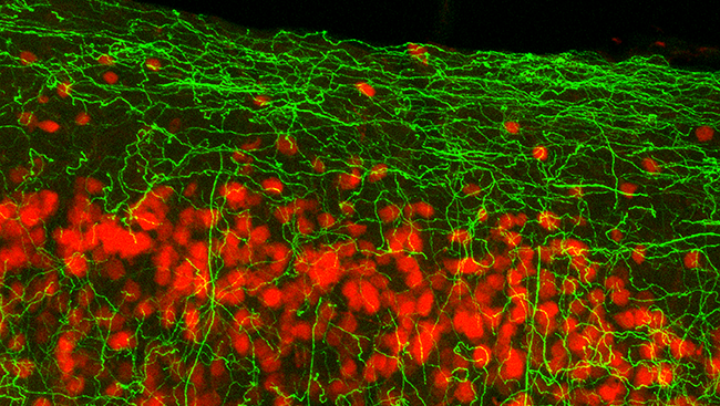
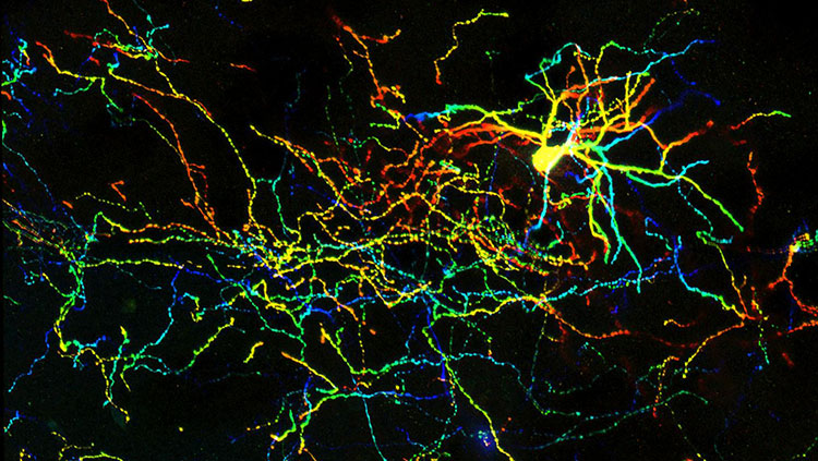
The cingulate cortex is a part of the limbic system, a collection of brain structures that plays a critical role in emotions, learning, and memory. This image shows the cingulate cortex in a mouse, with individual neurons stained in red. The basal forebrain, an area on the underside of the frontal lobe, sends nerve fibers (green) to regulate activity in the cingulate cortex. By mapping the location of these nerve fibers, scientists learned how they were organized, a discovery that could provide important insight into cognition and behavior.
About the Author

Alexis is a former staff writer/editor for BrainFacts.org. She graduated from the University of Pittsburgh in 2012 with degrees in neuroscience and English.
CONTENT PROVIDED BY
BrainFacts/SfN