The Molecules That Maintain the Brain
- Published6 Nov 2013
- Reviewed6 Nov 2013
- Author Mary Bates
- Source BrainFacts/SfN
We're all born with many more nerve cells than we eventually keep. Why do some cells survive and build intricate connections while others wither and die? How do nerve cells know which connections to make? The identification of a protein that tells nerve cells where to grow forever changed the way scientists think about cell growth and degeneration. Such insight is giving scientists new ways to study diseases such as Alzheimer’s disease and depression.
From perseverance to progress
The discovery more than 60 years ago of a protein that directs the development, survival, and function of nerve cells is giving scientists a new way to study and understand disorders of nerve cell growth — such as cancer — and degeneration — such as Alzheimer’s disease.
A determined quest for knowledge
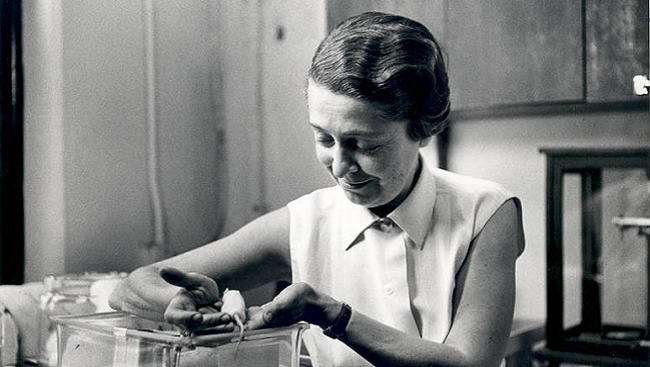
During the late 1930s in Italy, a young Jewish scientist by the name of Rita Levi-Montalcini found herself suddenly banned from the lab where she had spent years studying how nerve cells grow. Not to be deterred by the laws that forced those of Jewish descent from academia, Levi-Montalcini set up a makeshift lab where she began to secretly study chick embryos in search of an answer to a question that had been puzzling scientists for years: Early in development, how do nerve cells know where to go?
World War II forced Levi-Montalcini and her family to leave Turin for the countryside, where she continued her experiments in hiding. In 1947, after years of conducting her studies in secrecy, Levi-Montalcini joined scientists at Washington University in St. Louis, where they openly continued the quest to understand how the nervous system develops.
The Aha moment
In 1952, Levi-Montalcini discovered that transplanting pieces of mouse tumors to chick embryos stimulated the nerve cells of the chick to extend fibers in the direction of the tumors. Levi-Montalcini hypothesized the tumors were releasing a substance that guides nerve cell growth and promotes survival.
Years later, Levi-Montalcini, together with American biochemist Stanley Cohen isolated the substance directing nerve fibers to grow: a protein they named nerve growth factor (NGF). The work was monumental because it gave scientists a new way to study how the nervous system develops.
In 1986, Levi-Montalcini and Cohen received the Nobel Prize in Physiology or Medicine for their discovery of NGF.
Promoting brain cell survival, function
Scientists soon began to apply their knowledge of NGF — the protein cells release to lure new nerve fibers — to answer bigger questions of how nerve growth factors promote cell survival.
Guiding connections in the developing nervous system
Of the many nerve cells produced early in the development of the nervous system, only some will survive into adulthood. Because tissues only produce a limited amount of NGF, newborn nerve cells must compete for it in order to survive. Whether a cell lives or dies is determined by its ability to establish contact with the growth-promoting protein.
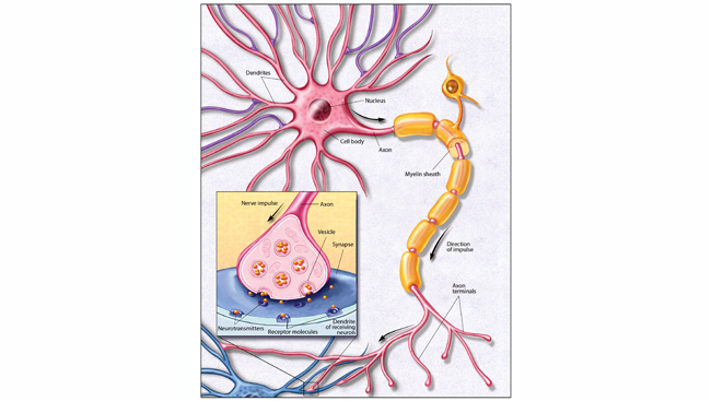
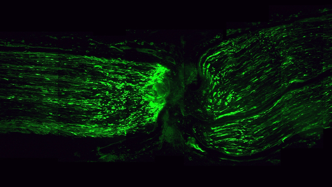
Similar to the way a plant grows toward sunlight, nerve cells extend their fibers in the direction of the NGF source. Once contact with the protein is made, it is absorbed by the end of the nerve, initiating a cascade of activity inside the cell. Such activity prompts the fibers to continue to extend, allowing, for instance, young nerves in the spinal cord to connect with developing limbs in early development. NGF also influences how cells connect and communicate with each other.
The role of brain nutrients throughout life
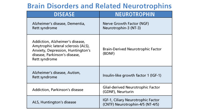
In the decades since the discovery of NGF, scientists have discovered many other growth-promoting proteins that act upon different types of cells in the nervous system. Scientists also continue to uncover new information about the changes that take place inside cells after these proteins, known collectively as neurotrophins, bind to their surfaces.
As it turns out, many neurotrophins are not only active in early development, but allow nerve cells to transmit signals with neighboring cells throughout life. For instance, numerous studies show NGF promotes the survival and communication of cells in the basal forebrain, a brain region that is involved in memory. Other studies show that the neurotrophin brain-derived neurotrophic factor (BDNF) helps the brain generate new neurons in the hippocampus, a memory center that receives information from the basal forebrain. Recent findings indicate these growth factors also strengthen connections between these brain cells.
Links to brain disorders and diseases
Because neurotrophins promote the health of the adult brain, scientists have become increasingly interested in how deficits in these proteins may be involved in neurodegenerative diseases and psychiatric disorders.
Low levels associated with disease
Human and animal studies have revealed the association between low levels of neurotrophins and brain diseases and disorders. For instance, one study found mice unable to process NGF as adults show memory loss and brain changes as they age that are characteristic of Alzheimer’s disease. Another shows people with depression or anxiety have reduced levels of BDNF.
There is also evidence that increasing neurotrophin levels may reverse some of the symptoms of these disorders. For instance, when scientists administered NGF to the mice showing brain changes similar to Alzheimer’s disease, brain function and behavior improved. In a separate study, scientists showed BDNF could halt and reverse age-related brain changes in mice, rats, and monkeys. Human studies show antidepressant medications, electroconvulsive therapy, and physical exercise all boost BDNF and can help ease symptoms of depression.
Overcoming challenges
These and other recent studies highlight the therapeutic potential of neurotrophins. However, there are challenges to translating the results into treatments.
Because neurotrophins are relatively large, they are unable to move easily across the blood-brain barrier — a network of blood vessels separating the brain from circulating blood. Additionally, delivering neurotrophins directly to the brain will require invasive and costly treatments. To get over these and other hurdles, scientists are working to find clues about the changes that take place inside a nerve cell after a neurotrophin binds to it.
Levi-Montalcini had no way of predicting just how great a role growth-promoting proteins have in brain health and disease when she refused to back away from her efforts to understand them in the late 1930s. However, her story demonstrates how commitments to basic science discovery, regardless of how trying the circumstances, can eventually open up a world of new information.
About the Author

Mary Bates is a freelance science writer interested in the brains and behavior of humans and other animals. For her graduate degree in psychology, she studied the echolocation abilities of big brown bats. She has written for Psychology Today, Scientific American's Mind Matters blog, the Howard Hughes Medical Institute, and other print and online publications.
CONTENT PROVIDED BY
BrainFacts/SfN
References
Allen SJ, Dawbarn D. Clinical relevance of the neurotrophins and their receptors. Clinical Science. 110: 175-191 (2006).
Arancio O, Chao MV. Neurotrophins, synaptic plasticity, and dementia. Current Opinion in Neurobiology. 17: 325-330 (2007).
Capsoni S, Ugolini G, Comparini A, Ruberti F, Berardi N, et al. Alzheimer-like neurodegeneration in aged antinerve growth factor transgenic mice. Proceedings of the National Academy of Sciences. 97(12): 6826–6831 (2000).
Castren E, Rantamaki T. The role of BDNF and its receptors in depression and antidepressant drug action: Reactivation of developmental plasticity. Developmental Neurobiology. 70(5): 289-297 (2010).
Chao MV. Neurotrophins and their receptors: A convergence point for many signaling pathways. Nature Reviews. 4: 299-309 (2003).
Chao MV, Rajagopal R, Lee FS. Neurotrophin signalling in health and disease. Clinical Science. 110: 167-173 (2006).
Chen KS, Nishimura MC, Armanini MP, Crowley C, Spencer SD, et al. Disruption of a single allele of the nerve growth factor gene results in atrophy of basal forebrain cholinergic neurons and memory deficits. Journal of Neuroscience. 17: 7288-7296 (1997).
Dawbarn D, Allen SJ. Neurotrophins and neurodegeneration. Neuropathology and Applied Neurobiology. 29: 211-230 (2003).
Leibrock J, Lottspeich F, Hohn A, Hofer M, Hengerer B, et al. Molecular cloning and expression of brain-derived neurotrophic factor. Nature. 341: 149-152 (1989).
Levi-Montalcini R. In Praise of Imperfection: My Life and Work. New York, 1988.
Nagahara AH, Merrill DA, Coppola G, Tsukada S, Schroeder BE, et al. Neuroprotective effects of brain-derived neurotrophic factor in rodent and primate models of Alzheimer's disease. Nature Medicine. 15(3): 331-337 (2009).
Shirayama Y, Chen A, Nakagawa S, Russell DS, Duman RS. Brain-derived neurotrophic factor produces antidepressant effects in behavioral models of depression. Journal of Neuroscience. 22(8): 3251-3261 (2002).
Tapia-Arancibia L, Aliaga E, Silhol M, Arancibia S. New insights into brain BDNF function in normal aging and Alzheimer disease. Brain Research Reviews. 59: 201-220 (2008).
Whitehouse PJ, Price DL, Struble RG, Clark AW, Coyle JT, et al. Alzheimer's disease and senile dementia: Loss of neurons in the basal forebrain. Science. 215(4537): 1237-1239 (1982).
Zuccato C, Cattaneo E. Brain-derived neurotrophic factor in neurodegenerative diseases. Nature Reviews Neurology. 5: 311-322 (2009).