Brain Scans: Technologies That Peer Inside Your Head
- Published26 Feb 2014
- Reviewed26 Feb 2014
- Author Aalok Mehta
- Source BrainFacts/SfN
The discovery that a chemist’s tool could create detailed internal images of the body more than 40 years ago set into motion a series of advances that forever changed how scientists study brain structure and function. Today, researchers use magnetic resonance imaging (MRI) to study the healthy and unhealthy brain. These and other emerging technologies may soon guide scientists to create an even more dynamic picture of the brain.
Physical scientists uncover way to see inside body
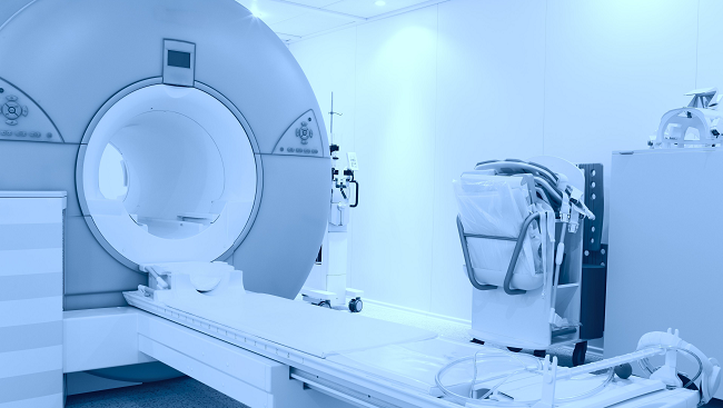
More than 60 million MRI scans take place around the world each year in both research and clinical settings. But MRI might have never come to be if it were not for a group of scientists rethinking how a technique they had used for years could be applied to the human body. The story of how MRI made its way to the clinic is one of many examples of the role that chemistry, physics, math, and computer science play in brain research and health.
A matter of chemistry
Chemist Paul Lauterbur was on summer break from teaching in the early 1970s when he was tapped to lead a struggling scientific company from the brink of bankruptcy. The company sold and leased nuclear magnetic resonance (NMR) spectrometers, a technology that chemists used to determine the identity of chemical compounds. While there, Lauterbur became increasingly interested in data that showed NMR could distinguish between samples of cancerous and healthy body tissue. He wondered if NMR could be used to recreate pictures of tissues and organs inside the living body.
Lauterbur knew that water content varies in different types of body tissue and that NMR could detect the hydrogen atoms in water molecules. But, at the time, NMR could not determine the position of the atoms. Lauterbur realized he could determine the position of atoms in a sample by introducing variations in the magnetic field. This information could then be used to build 3-D images reflecting the chemical structure of organs and tissues.
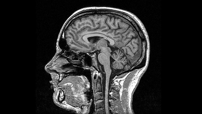
Using mathematical techniques, the British physicist Peter Mansfield dramatically reduced the time it took to collect NMR information and transform the information into an image using advanced computers. The resulting black-and-white image revealed tissues high in water and fat content in white and those low in water and fat in black.
In the early 1980s, this new imaging technology made its debut in hospitals as MRI, and with it, the study of the brain and nervous system forever changed.
MRI illuminates brain structure, function
MRI offered physicians several advantages over the other imaging techniques of the 1980s. In addition to producing high-quality, 3-D images of organs and tissues inside the body, MRI did not require that a patient be exposed to radiation, as is the case with X-rays and computed tomography (CT) scans.
The ability to visualize organs and tissues made it easier for doctors to diagnose, track, and treat brain and spinal cord abnormalities resulting from disease and injury. However, it was the revelation that MRI could track changes in brain activity and the connections between nerve cells that revolutionized brain science.
A flurry of activity
Multiple brain areas are hard at work at any given time. As a brain region becomes more active, it consumes more oxygen. In response, the body rushes in oxygen-rich blood to the active area. In the 1990s, scientists realized that the magnetic properties of oxygen-rich and oxygen-poor blood differ, and these differences could be measured by a variation of MRI called functional MRI (fMRI).
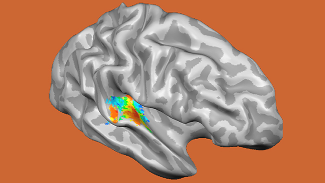
Unlike other techniques, fMRI offered a way for scientists to noninvasively measure changes in activity deep inside the brain. With this new ability, researchers could begin to see how the brain changes as we look at a picture of a loved one or perform a simple task.
Key connections
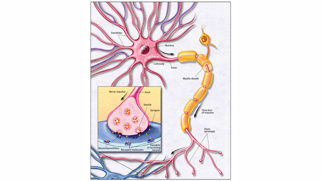
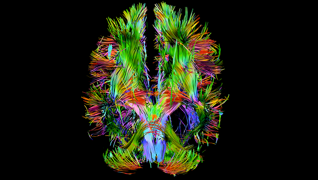
With traditional MRI, scientists could see nerve cell bodies (similar to the major landmarks on a map) but not the nerve fiber connections between them called axons (similar to the highways linking regions). The development of diffusion tensor imaging (DTI), another variation of MRI, gave scientists the ability to visualize these connections. DTI tracks the preferential movement of water molecules along the axons connecting cells, revealing the pathways of the brain. DTI studies have led to greater knowledge about how the brain continues to change throughout life.
Imaging the brain in health and disease
With better techniques to study brain activity and connections, scientists have been able to ask big and answer big questions about brain structure and function. These answers and resulting new questions are propelling the field into a new era of learning about brain health and disease.
The busy brain
Using fMRI, scientists can now pinpoint the brain regions most active when we perform a variety of tasks — from recognizing faces to contemplating moral responsibility. fMRI studies also show the brain remains active even when people are asleep or under anesthesia.
DTI continues to reveal new information about how the brain changes throughout life. It has also proven useful for tracking short- and long-term structural damage caused by injuries such as concussions, allowing physicians to assess brain damage long after symptoms resolve.
While these studies are advancing knowledge of brain structure and function, scientists are careful to note there is still much to understand about how such information translates to cognitive function.
The big picture
Just as MRI transformed the diagnosis of cancer, scientists hope the next generation of imaging techniques might aid in the early diagnosis of neurodegenerative diseases such as Alzheimer’s and Parkinson’s, and psychiatric disorders, including depression and schizophrenia.
Even as researchers look for new ways to apply current imaging technology, many have their sights set on the creation of an even more dynamic picture of the brain. The creation of new tools to study the brain has become one of the most pressing needs and exciting opportunities for today’s neuroscience field. Large-scale projects funded by public and private entities in the United States and Europe aim to further advance imaging technology and other technologies that collect, store, distribute, share, and analyze data on brain anatomy and activity. Such advances will help to fill in current knowledge gaps regarding how the brain works, advancing science and forming the foundation for new health advances.
CONTENT PROVIDED BY
BrainFacts/SfN