ICYMI: One Distinct Neuron Population Dwindles in Parkinson’s Disease
- Published17 Jun 2022
- Author Tristan Rivera
- Source BrainFacts/SfN

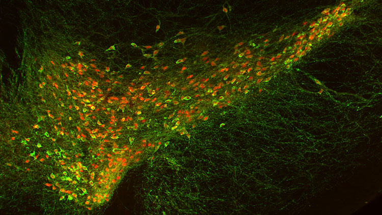
The ability to zoom in and study compact areas of the brain allows us to better understand the microscopic world contained within. Recently, this view has helped identify specific brain cells that die off in Parkinson’s disease (PD). PD results from the loss of dopamine-producing cells in the substantia nigra, an area within the midbrain that manufactures dopamine and influences aspects of movement control, higher-level cognition, and our emotions. New research published May 5 in Nature Neuroscience parses apart a particular dopamine-making cell population in the substantia nigra that is drastically diminished in people with PD — providing a major insight into specific targets for future treatments of the disease.
Researchers from the Broad Institute of Harvard and MIT looked at the postmortem brains of eight people with no evidence of neurodegeneration and 10 people with documented neuron loss in the midbrain (associated with PD and Lewy body dementia). Using single-nucleus RNA-sequencing, researchers viewed which genes were turned on, or expressed, in individual cells by isolating their nuclei. The researchers then visualized large swaths of gene expression patterns across the substantia nigra with fine precision.
Researchers later identified 10 groups of dopamine-making neuron subpopulations in the substantia nigra. They found high susceptibility of neuron loss in one specific subpopulation located on the underside of the substantia nigra, marked by activation of a gene called AGTR1. The study not only provided an enhanced topographic view of the substantia nigra, but also a clue into what specific area of the substantia nigra illuminates the process of PD-related degeneration.
Big Picture: First identified in 1817, PD has been long studied. In the late 1950s, the discovery of decreased dopamine levels in PD patients helped spur the 1960s advent of levodopa, a drug that helps replace dopamine in the brain. PD treatment breakthroughs such as dopamine replacement therapy, deep brain stimulation, and gene therapy have since made significant gains at enhancing the lives of those afflicted by PD. The next step — isolating which brain regions are most susceptible to PD — can help orient researchers toward more targeted studies and treatments.
Read more: A very specific kind of brain cell dies off in people with Parkinson’s. Science News
More Top Stories
- Seizures can induce myelination in rodents, enhancing the efficiency of neuronal firing and contributing to epilepsy progression. STAT News
- A test combining memorization and changing pupil size may help determine whether people have aphantasia, the inability to picture images in their head. New Scientist
- Early inflammation may provide a protective effect to avoid chronic lower-back pain. STAT News
- Researchers are attempting to categorize what we know about animal intelligence. New Scientist
- Our fascia tissue surrounds our muscles and organs — but recent findings have some debating if it should be considered a sensory organ. New Scientist
- The Protactile movement provides DeafBlind communities the opportunity to communicate through touch. The New Yorker
- Boosted production of a steroid may be why octopuses starve themselves after mating. New Scientist
- An infusion of spinal fluid from young mice helps old mice better perform memory tasks. NPR
- Why contagious yawns occur — and why they aren’t specific to humans. Science
CONTENT PROVIDED BY
BrainFacts/SfN
References
History of PD. (n.d.). Stanford Parkinson’s Community Outreach. Stanford Medicine. Accessed June 15, 2022. https://med.stanford.edu/parkinsons/introduction-PD/history.html
Kamath, T., Abdulraouf, A., Burris, S.J. et al. (2022). Single-cell genomic profiling of human dopamine neurons identifies a population that selectively degenerates in Parkinson’s disease. Nat Neurosci 25, 588–595. https://doi.org/10.1038/s41593-022-01061-1
Rodriques, S.G., Stickels, R.R., Goeva, A., Martin, C.A., Murray, E., Vanderburg, C.R., Welch, J., Chen, L.M., Chen, F., Macosko, E.Z. (2019). Slide-seq: A scalable technology for measuring genome-wide expression at high spatial resolution. Science 363, 1463-1467. https://doi.org/10.1126/science.aaw1219
Sonne, J., Reddy, V., Beato, M. (2021). Neuroanatomy, Substantia Nigra. StatPearls [Internet]. Accessed June 17, 2022. https://www.ncbi.nlm.nih.gov/books/NBK536995/
Wasinski, F., Pedroso, J.A.B., Santos, W.O., Furigo, I.C., Garcia-Galiano, D., Elias, C.F., List, E.O., Kopchick, J.J., Szawka, R.E., Donato, J. (May 2020). Tyrosine Hydroxylase Neurons Regulate Growth Hormone Secretion via Short-Loop Negative Feedback [Journal cover image]. Journal of Neuroscience 40 (22) 4309-4322, Figure 9D. https://doi.org/10.1523/JNEUROSCI.2531-19.2020


