Transcranial Magnetic Stimulation Can Improve Visual Perception
- Published12 Apr 2022
- Author Jordan Anderson
- Source BrainFacts/SfN
You’re about to cross the street when you spot a car barreling toward the intersection. A radiologist reviewing a patient's scan must distinguish healthy tissue from a tumor.
In both scenarios, fast and accurate visual perception is vital. It’s a skill we can improve with practice. For example, a 2020 study demonstrated playing Where’s Waldo helped radiology residents spot abnormalities on patient x-rays.
But it may be possible to improve perception without all that hard work. In a small, proof-of-concept study, researchers found stimulating the brain with magnets helped people perceive objects faster. The findings, which have yet to be published, were presented at a press conference at the annual meeting of the Society for Neuroscience in November 2021.
“This is an important technical accomplishment,” Colleen Hanlon, a neurobiologist at Wake Forest University School of Medicine who studies brain stimulation, said in an email. She was not involved in the study. It “adds to an emerging body of literature supporting the feasibility of online, closed loop brain stimulation as a tool to enhance attention and human performance.”
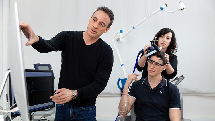
Transcranial magnetic stimulation (TMS) stimulates brain cells using magnetic waves. The non-invasive technique has been used to treat depression in people who don’t respond to medication. In the last three decades, scientists have found that stimulating specific brain regions can enhance brain and cognitive function.
Nitzan Censor and his colleagues at Tel Aviv University wanted to know if visual perception could be enhanced by closed-loop stimulation of remote brain regions. Closed-loop stimulation uses real-time neuroimaging to continuously monitor the brain state of a particular region. This monitoring system provides feedback, adjusting stimulation to reach a desired brain state. When the stimulation alters the brain state of the monitored region, it is considered ‘closed-loop’ — much like a thermostat monitors temperature and sets off a trigger to bring a room to a desired temperature.
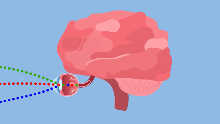
Visual information travels from the eyes to the visual cortex. Then there is a back-and-forth flow of information between the visual cortex and the frontal cortex that helps us make sense of what we see. “If we can increase the connectivity between these regions, perhaps this will [also] enhance visual perception,” Censor says.
He and his team tested the perception of ten volunteers with and without TMS. Participants looked at a computer screen where small, hard-to-see spots appeared for a few milliseconds before disappearing. They pressed a keyboard to indicate when they saw each spot. During one test session, Censor and his colleagues applied TMS to the top left of each participant’s head, over the right frontal eye field. Censor chose this area because it communicates with the visual cortex and helps us focus our visual attention. Participants detected the spots more accurately when TMS was applied.
At the same time, Censor also recorded from participants’ occipital lobes using electroencephalography (EEG). They used the signals recorded in the occipital lobe to guide the timing of stimulation delivered to the frontal lobe. This closed-loop stimulation helped facilitate communication between the two regions as they applied TMS stimulation when the occipital lobe was ready to exchange information.
“While TMS has been widely tested to enhance brain function and its corresponding behavior, the core novelty [in this experiment] is to enhance perception by improving inter-regional brain connectivity,” Censor said in an email.
Using TMS for superspeed vision is far from becoming a reality. The benefits of the technique are short lived, and they don’t replace initial learning. A radiologist, for example, still has to learn what normal and abnormal x-rays look like before they can diagnose their patients. Still, the findings open exciting possibilities. To Hanlon, one of the most intriguing aspects of the study is that it capitalizes on “the role of the frontal eye field in attention.” “It will be very interesting to see if this can be applied as a therapeutic approach to clinical populations that suffer from disorders of attention,” she says.
CONTENT PROVIDED BY
BrainFacts/SfN
References
Bergmann, T. O. (2018). Brain State-Dependent Brain Stimulation. Frontiers in Psychology, 9. https://www.frontiersin.org/article/10.3389/fpsyg.2018.02108
Carbon, C.-C. (2014). Understanding human perception by human-made illusions. Frontiers in Human Neuroscience, 8. https://www.frontiersin.org/article/10.3389/fnhum.2014.00566
Mulckhuyse, M., Kelley, T. A., Theeuwes, J., Walsh, V., & Lavie, N. (2011). Enhanced visual perception with occipital transcranial magnetic stimulation. The European Journal of Neuroscience, 34(8), 1320–1325. https://doi.org/10.1111/j.1460-9568.2011.07814.x
Noohi, S., & Amirsalari, S. (2016). History, Studies and Specific Uses of Repetitive Transcranial Magnetic Stimulation (rTMS) in Treating Epilepsy. Iranian Journal of Child Neurology, 10(1), 1–8.
Sahraian, S., Yousem, D., Beheshtian, E., Jalilianhasanpour, R., Morales, R. E., Krupinski, E. A., & Zhan, H. (2020). Improving Radiology Trainees’ Perception Using Where’s Waldo? Academic Radiology, S1076-6332(20)30611-5. https://doi.org/10.1016/j.acra.2020.10.023
Transcranial Magnetic Stimulation (TMS) Service. (n.d.). Johns Hopkins Medicine. Retrieved March 28, 2022, from https://www.hopkinsmedicine.org/psychiatry/specialty_areas/brain_stimulation/tms/


