
Synopsis
In this activity, students elicit and observe reflex responses and distinguish between types of reflexes. They then design and conduct experiments to learn more about reflexes and their control by the nervous system.
Estimated time needed: 1–2 class periods.
Objectives
By the end of this lesson, students will be able to:
- Identify common reflexes of the human body and the neural circuitry involved
- Distinguish between stretch and protective reflexes
- Describe the role of reflexes in the body
Subjects
- Biology, Physics, Anatomy & Physiology
Materials
For each group of 12 students:
- 1 rubber percussion hammer or 30-cm (12-in) ruler
- 1 flashlight or penlight
- 1 pair safety goggles
- 1 square of light cardboard (5 × 5 cm or 2 x 2 in)
- 1 marker
Background: Knee-Jerk and Pupillary Light Reflexes
This activity will provide students an opportunity to see the knee-jerk (patellar) and pupillary light reflex in action. Before beginning the activity, students should have a general understanding of how the nervous system detects stimuli and produces rapid responses. Students can read “Understanding Involuntary Movements” on BrainFacts, which explains how the body and spinal cord dictate certain movements, like reflexes, without our conscious control.
The knee-jerk reflex is an automatic response that helps keep us balanced and standing upright. When the tendon just below the kneecap is tapped, it briefly stretches the thigh muscle. Sensory neurons send a signal to the spinal cord, which quickly sends a message back to the muscle, causing the lower leg to kick forward. This reflex happens without conscious thought and is one of the fastest responses in the nervous system.
The pupillary light reflex helps regulate the amount of light entering the eyes, protecting the eyes and improving vision in different lighting conditions. When bright light enters the eye, sensory cells in the retina detect it and send signals to the brain. The brain then sends signals to the muscles of the iris, causing the pupil to become smaller. This limits the amount of light entering the eye. When light levels decrease, the pupils become larger again to let in more light.
Exploration Activity: Part One — Knee-Jerk Reflex
- Divide the class into groups of four students and have each group conduct the activity. Assign each student one of the following roles: subject, experimenter, timer, and data recorder.
- Have each group do the knee-jerk reflex. The subject should relax and sit on a desk or table with his or her legs dangling loosely over the edge.
- The experimenter should apply the stimulus by tapping, firmly but carefully, the area just below the kneecap with a percussion hammer or ruler.
- The data recorder should indicate to the timer when the experimenter applies the stimulus, and the timer should note the time.
- The data recorder should indicate to the timer when the subject has responded, describe the response, and record the time.
- The group should repeat these steps with the other leg.
Exploration Activity: Part Two — Pupillary Light Reflex
- Have students exchange roles and conduct the pupillary light reflex.
- In a dimly lit room, the subject should look toward a wall until his or her eyes dilate. The experimenter should place a square of light cardboard on the bridge of the subject’s nose to separate each eye’s field of vision.
- The experimenter should bring a flashlight to within 5–10 cm (2–5 in) of the subject’s face. The timer should signal the experimenter to turn on the light for 3–5 seconds.
a. Safety Note: Do not shine the flashlight directly into anyone's eyes for longer than 3–5 seconds. Use only a standard classroom or household flashlight — not a high-intensity LED, laser pointer, or other bright light source. If the subject experiences discomfort, sensitivity to light, or changes in vision, stop the activity immediately. Keep the flashlight 5–10 cm (2–5 in) from the face and avoid physical contact with the eye. - The data recorder should observe the response of the pupil in the eye that is stimulated and the one that is not and record the results.
- Repeat the test with the other eye and note the response in both eyes.
Lesson Wrap-Up: What’s Happening?
A reflex is an automatic, involuntary response to a sensory stimulus. Reflexes help the body respond quickly to changes in the environment and play important roles in maintaining balance, posture, vision, and overall homeostasis.
Throughout this activity, students observed two examples of reflexes in action: the knee-jerk reflex and the pupillary light reflex.
Both the knee-jerk reflex and the pupillary light reflex demonstrate how the nervous system can respond rapidly without conscious effort. Although they each produce different responses, they follow a similar pattern. The knee-jerk reflex uses spinal cord circuits to help maintain posture and balance, while the pupillary light reflex uses brainstem pathways to regulate the amount of light entering the eyes. Together, these reflexes show how different parts of the nervous system work continuously to keep the body functioning safely and efficiently.
How the Knee-Jerk (Patellar) Reflex Works
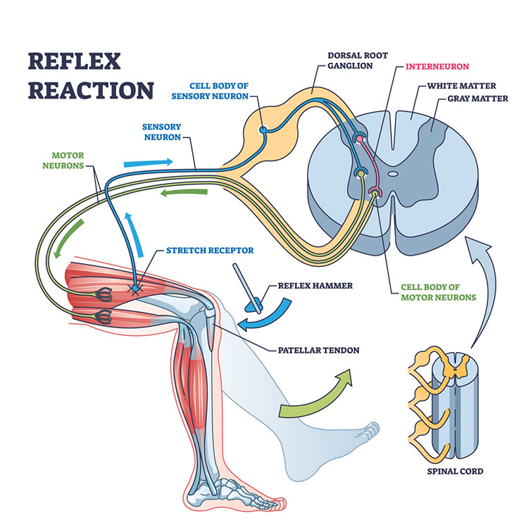
When the patellar tendon was tapped during the activity, the quadriceps muscle was briefly stretched. Specialized stretch receptors detected the change and sent a signal through a sensory neuron to the spinal cord. The cell body of the sensory neuron is located in the dorsal root ganglion, just outside the spinal cord.
Within the spinal cord's gray matter, the sensory neuron directly activates the cell bodies of motor neurons, making the knee-jerk reflex one of the body's simplest and fastest reflexes. The sensory neuron also communicates with an interneuron, which inhibits motor neurons controlling opposing muscles so the leg can move smoothly. Signals then travel back through motor neurons to the quadriceps muscle, causing the lower leg to kick forward.
The spinal cord contains both gray matter, where neurons communicate, and white matter, which contains nerve fibers that carry information throughout the nervous system. Because the reflex is processed primarily in the spinal cord rather than waiting for instructions from the brain, the response occurs automatically and very quickly. This helps the body maintain posture, balance, and muscle stability during movement.
How the Pupillary Light Reflex Works (Overview)
The pupillary light reflex is a brainstem reflex that automatically adjusts pupil size in response to light.
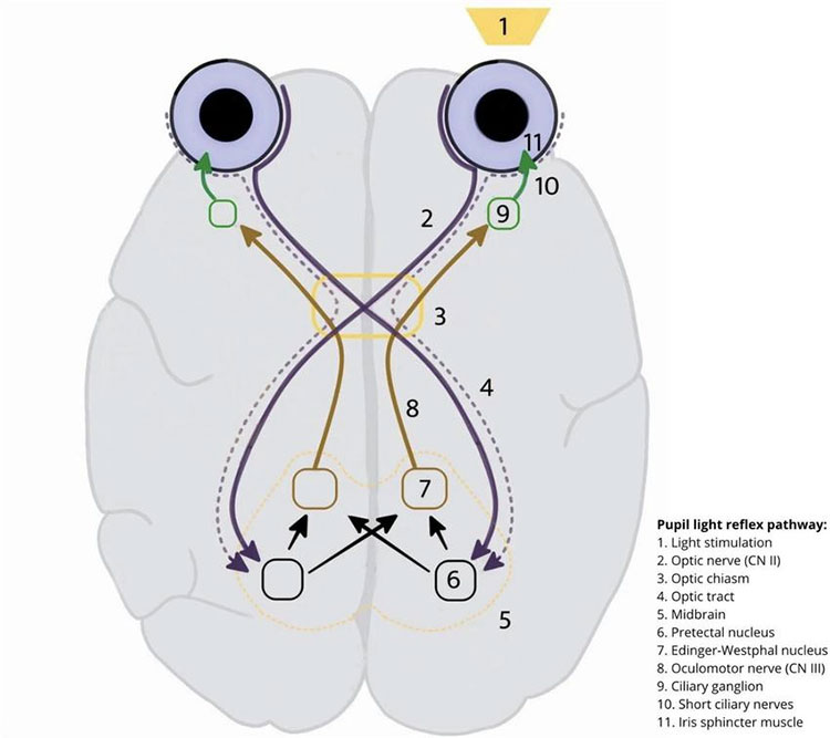
During this activity, students observed what happened when a flashlight shined into one eye in a dim room. Before the light was turned on, the pupils were dilated to allow more light into the eyes. When light entered one eye, students observed the pupil in that eye to become smaller. They may also have noticed that the pupil in the opposite eye became smaller at the same time. These responses are known as the direct response and consensual response, respectively.
The pupillary light reflex helps regulate how much light enters the eyes. Light information travels from the eye to the brain and back to the eye through a series of nerve pathways, as indicated in Figure 2. Because signals are sent to both sides of the brainstem, shining light into one eye normally causes both pupils to constrict. This reflex helps protect the retina and maintain clear vision in changing light conditions.
(Optional) Deeper Dive: Pupillary Light Reflex
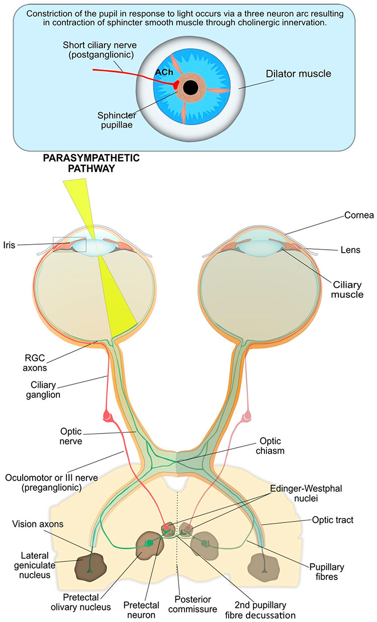
As indicated in Figure 3, the reflex begins when light passes through the cornea and lens and reaches the retina. Specialized retinal ganglion cells generate signals that travel along the optic nerve, through the optic chiasm, and into the optic tract. Some of these fibers branch away from the visual pathway and travel to the pretectal olivary nucleus in the midbrain. Pretectal neurons then send signals to both Edinger–Westphal nuclei, crossing through the posterior commissure and creating a second crossing of pupillary fibers. This bilateral pathway ensures that information from one eye reaches both sides of the brain.
From the Edinger–Westphal nuclei, signals travel through the oculomotor (III) nerve to the ciliary ganglion. The signal then continues through the short ciliary nerves, which release acetylcholine (ACh) onto the sphincter pupillae muscle of the iris. As the sphincter pupillae contracts, the pupil becomes smaller. At the same time, the opposing dilator muscle becomes less active. The result is a rapid decrease in pupil size that protects the retina from excessive light and helps optimize vision under changing light conditions.
Because signals are sent to both Edinger–Westphal nuclei, illuminating one eye normally causes both pupils to constrict, allowing the visual system to respond quickly and automatically to changes in brightness.
CONTENT PROVIDED BY
BrainFacts/SfN
Discussion Questions
-
How did your body respond to the stimulus? How quickly did it respond? Can you control your reaction?
-
What function do these responses serve? Why are they important for survival?
-
How are the stretch reflex and the pupillary light reflex similar and different?
References
Falkson S. R., Hinson J. W. Westphal Sign. [Updated 2023 Feb 6]. In: StatPearls [Internet]. Treasure Island (FL): StatPearls Publishing; 2026 Jan-. Available from: https://www.ncbi.nlm.nih.gov/books/NBK553214/
Hall, C. A., & Chilcott, R. P. (2018). Eyeing up the Future of the Pupillary Light Reflex in Neurodiagnostics. Diagnostics, 8(1), 19. https://doi.org/10.3390/diagnostics8010019
Lisowski, Ł., Lisowska, J., Charytonowicz, A., Mariak, Z., Obuchowska, I., & Konopińska, J. (2025). The diagnostic significance of pupillary reflex pathways: Insights from classical examination and advanced pupillometry. Frontiers in Neuroscience, 19, Article 1677431. https://doi.org/10.3389/fnins.2025.1677431